Checking Out the Necessary Providers Supplied by a Vet Cardiologist: Comprehending Ultrasound and CT Scan Methods
Veterinary cardiologists play an important role in the health of pets by detecting and dealing with different heart disease. They utilize advanced imaging techniques, such as heart ultrasound and CT scans, to give exact examinations. Each technique has its distinct advantages and applications. Recognizing these methods is vital for animal owners looking for the very best take care of their friends. What aspects should animal proprietors think about when selecting in between these diagnostic devices?

The Function of Vet Cardiologists in Pet Healthcare
Veterinary cardiologists play an essential function in the medical care of pet dogs, concentrating specifically on diagnosing and treating heart-related conditions. They possess specialized training that allows them to translate complicated diagnostic tests and determine different cardiovascular problems. These experts use sophisticated methods, such as echocardiography and electrocardiography, to examine heart feature and structure accurately.Veterinary cardiologists likewise establish tailored treatment strategies that might consist of drugs, way of living adjustments, and, in many cases, medical interventions. Their competence includes informing animal owners about heart wellness, stressing the relevance of normal exams and early discovery of possible troubles. Partnership with general veterinarians is vital, as it assures detailed treatment for animals with suspected heart problems. By providing specialized services, veterinary cardiologists greatly enhance the top quality of life for animals and offer comfort for their owners, reinforcing the importance of heart wellness in total family pet wellness.
Common Cardiac Concerns in Pets
Usual cardiac problems in pets can greatly influence their wellness and lifestyle. Heart whisperings, different sorts of cardiomyopathy, and hereditary heart defects are among the most prevalent problems that veterinarians experience. Ultrasound For Dogs. Comprehending these issues is important for pet proprietors to assure timely diagnosis and suitable therapy
Heart Murmurs in Pets
Heart murmurs can be a resource of concern for family pet owners, they are not always indicative of serious wellness issues. A heart whispering is an unusual noise generated by turbulent blood flow within the heart. In family pets, these whisperings can be brought on by various elements, consisting of genetic heart issues, valve issues, and even stress during evaluations. Lots of pet dogs with heart whisperings lead regular lives without significant health impacts. To identify the underlying cause, veterinary cardiologists commonly use analysis strategies such as echocardiograms and Doppler ultrasounds. Early discovery and analysis are crucial, as they may help manage any type of prospective heart concerns properly. Pet owners are encouraged to consult their veterinarian for a detailed analysis if a heart whispering is identified.
Cardiomyopathy Types Explained
Cardiomyopathy encompasses a group of conditions influencing the heart muscle, leading to compromised cardiac function in pet dogs. The most typical types consist of expanded cardiomyopathy (DCM), hypertrophic cardiomyopathy (HCM), and limiting cardiomyopathy (RCM) DCM primarily influences dogs, causing the heart to enlarge and deteriorate, which lessens its ability to pump blood effectively. In contrast, HCM is much more common in felines, defined by the thickening of the heart walls, typically leading to blocked blood circulation. RCM, though less usual, occurs when the heart muscle mass comes to be inflexible, limiting its ability to fill with blood. Each type presents unique difficulties in medical diagnosis and therapy, requiring specialized veterinary cardiological examination to assure peak management and take care of influenced family pets.
Congenital Heart Defects
Hereditary heart flaws stand for a considerable group of heart problems in animals, unique from obtained conditions such as cardiomyopathy - CT Scans For Animals. These defects are architectural irregularities present at birth, affecting the heart's regular feature. Common kinds include patent ductus arteriosus, ventricular septal flaws, and pulmonic stenosis. Signs might differ widely, varying from moderate to extreme, and can consist of exercise intolerance, coughing, and trouble breathing. Early diagnosis through advanced imaging methods like ultrasound is necessary for efficient management. Veterinary cardiologists play a crucial duty in identifying these conditions and suggesting ideal treatment options, which may consist of clinical monitoring or surgical intervention. Recognizing hereditary heart flaws enables better outcomes and enhanced high quality of life for influenced pet dogs
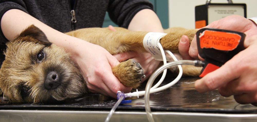
Understanding Cardiac Ultrasound: How It Works
A significant number of veterinary practices now use cardiac ultrasound as a necessary analysis device for assessing heart health and wellness in animals. This non-invasive method utilizes high-frequency acoustic waves to produce photos of the heart's framework and function. During the procedure, a veterinary technician applies a gel to the pet's upper body and makes use of a transducer to produce ultrasound waves. These waves bounce off the heart and surrounding structures, generating real-time photos on a monitor.Veterinarians can analyze numerous facets of heart health, consisting of chamber dimension, wall surface activity, and shutoff function. Furthermore, heart ultrasound enables the detection of problems such as liquid buildup and genetic heart defects. This technique is vital for diagnosing problems that might not be visible through basic radiographs. By providing detailed details concerning the heart's composition and performance, heart ultrasound aids in developing effective therapy strategies for pets struggling with heart condition.
The Relevance of CT Scans in Diagnosing Heart Issues
How do CT scans enhance the medical diagnosis of heart disease in vet medicine? CT scans offer detailed cross-sectional pictures of the heart and surrounding frameworks, allowing vets to picture complex physiological relationships. This imaging method is particularly helpful in determining genetic heart problems, heart growths, and problems in blood vessels. By utilizing innovative imaging formulas, CT scans can analyze heart chamber dimensions and function, supplying a complete view that may be difficult to attain with standard methods.Additionally, CT angiography can picture blood circulation and identify areas of constriction or obstruction, which is essential for preparing possible interventions. The speed and precision of CT scans additionally facilitate quick medical diagnoses, essential in emergency circumstances. Inevitably, the incorporation of CT scans right into vet cardiology greatly improves the precision of medical diagnoses, making it possible for targeted treatment plans and improving individual end results for pets struggling with heart conditions.
Comparing Ultrasound and CT Scan Methods
While both ultrasound and CT scans are very useful devices in veterinary cardiology, they provide unique advantages and restrictions that affect their usage in detecting heart conditions. Ultrasound, or echocardiography, offers real-time imaging of the heart's framework and feature, permitting vets to assess heart chambers, valves, and blood flow. It is specifically reliable for evaluating conditions like coronary infarction and cardiomyopathy. Ultrasound might be restricted in envisioning particular physiological frameworks due important site to person dimension or obesity.In comparison, CT scans offer detailed cross-sectional photos of the heart and bordering tissues, making them perfect for identifying architectural irregularities, tumors, or vascular issues. CT scans provide thorough understandings, they need sedation and might include radiation exposure. Inevitably, the option between ultrasound and CT checks depends on the details scientific scenario, the person's condition, and the details needed for an exact medical diagnosis.
Treatment Choices Available Via Veterinary Cardiology
Veterinary cardiology offers an array of treatment alternatives tailored to resolve different heart disease in pets. Treatment strategies commonly begin with way of living alterations, including diet regimen changes and workout modifications, focused on enhancing total heart health and wellness. Drugs play an essential function, with cardiologists prescribing medications such as diuretics, beta-blockers, and ACE preventions to enhance and handle signs and symptoms cardiac function.In a lot more extreme cases, interventional treatments, such as balloon valvuloplasty or stent placement, may be essential to minimize clogs or improve blood flow. For specific genetic heart defects, surgical choices might be discovered to correct structural problems. Furthermore, recurring surveillance and follow-up care are vital parts of a comprehensive treatment plan, enabling for prompt changes based upon the pet dog's response to therapy. In general, veterinary cardiology concentrates on offering efficient, personalized treatment to enhance the health and wellness and health of pet additional info people with heart disease.
Just how to Prepare Your Pet Dog for a Heart Analysis
Preparing a pet dog for a heart analysis is necessary to guarantee exact outcomes and a smooth procedure. Proprietors ought to first schedule the appointment with the vet cardiologist and review any kind of details needs or worries. It is a good idea to keep food for at least 12 hours prior to the evaluation, as this aids improve imaging high quality during procedures like ultrasound or CT scans.Additionally, maintaining a tranquil setting on the day of the visit can help in reducing the pet dog's anxiousness. It is helpful to bring along any type of relevant clinical documents, including previous tests and drugs (CT Scans For Dogs). Owners need to likewise make certain that their pet dog fits and leashed during transportation to the center. Ultimately, acquainting themselves with the examination process can help and minimize worries in asking informed inquiries throughout the consultation. By adhering to these actions, proprietors can contribute greatly to the efficiency of the heart examination
Regularly Asked Questions
Just how Long Does a Cardiac Ultrasound or CT Check Take?
The period of a cardiac ultrasound commonly ranges from 30 to 60 minutes, while a CT check may take around 15 to thirty minutes. Elements such as the person's learn the facts here now condition can influence these time price quotes.

Exist Any Risks Associated With These Diagnostic Procedures?
Can I Keep With My Pet Dog Throughout the Procedure?
The veterinary facility's plan generally dictates whether animal owners can continue to be throughout treatments. While some facilities urge owner presence for comfort, others might need separation to guarantee safety and excellent conditions for diagnostic imaging.
Just how Much Do These Analysis Tests Typically Price?
The expenses of diagnostic examinations, such as ultrasound and CT scans, normally vary based upon place and facility. Commonly, rates range from a couple of hundred to over a thousand dollars, reflecting the intricacy and technology included.
What Is the Recovery Refine After a Cardiac Analysis?
The healing procedure after a heart evaluation entails keeping track of the pet dog for any type of immediate responses, making certain comfort, and restricting exercise. Vets usually provide post-evaluation directions to guide family pet owners during this necessary recovery duration. Heart whisperings, various kinds of cardiomyopathy, and genetic heart problems are amongst the most widespread conditions that vets encounter. A heart murmur is an irregular sound created by stormy blood flow within the heart. Cardiomyopathy includes a team of conditions impacting the heart muscular tissue, leading to compromised cardiac feature in pets. Congenital heart problems stand for a substantial category of cardiac problems in family pets, distinctive from acquired problems such as cardiomyopathy. Ultrasound, or echocardiography, provides real-time imaging of the heart's structure and feature, allowing vets to evaluate heart chambers, valves, and blood flow.